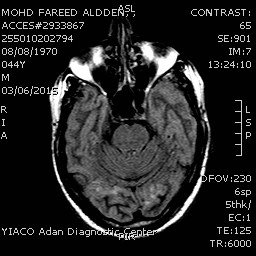


PROTOCOL:
T2W – Sagittal & axial scans
T1W – Axial scans
FLAIR – Coronal scans
T2FFE (Gradient) – Axial scans
3D TOF MR angiography of the circle of Willis vessels
and MIP reconstruction.
FINDINGS:
-REVEALED:
·
Bilateral
occipital areas of altered signal with faint hyper intensity on Flair images
cortical and sub cortical.
·
Abnormal
enhancement in the previously mentioned areas with abnormal meningeal
enhancement.
-Brain Parenchyma shows normal grey white matter
differentiation and signal intensities.
No focal mass lesion is seen.
-Lateral and third ventricles are normal in size and
position. Septum is in midline. There is
no midline shift.
-Basal cisterns, cortical sulci and gyri are normal in
appearance.
-Corpus callosum shows normal MR morphology.
-Brain stem and cerebellum show normal signal
intensities.
-Fourth ventricle is normal in size and is midline in
position.
-MR angiography reveals normal course, caliber and
outlines of vessels of bilateral CA, ACA, MCA, PCA and their peripheral
branches. Vertebro basilar trunk is also normal in appearance.
OPINION:
·
Picture is suggestive of cortical and
meningeal infection for clinical correlation.
Reported By : DR.MOHAMMED
MOHYELDIN
Consultant-Radiologist
No comments:
Post a Comment