


PROTOCOL:
T1W – Sagittal & axial scans
T2W – Axial & coronal scans
FLAIR – Axial scans
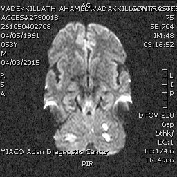
FINDINGS:
-Revealed:
• Multiple variable sized foci of altered MR signals in both cerebellar hemispheres with the largest one diameter 22 x 12 mm on the right side.
• Another focus in the left thalamus and in the left occipital region.
• Small vessel ischemic disease with grade I Fazekas classification.
• Non visualization of the left vertebral artery (occluded) on MR angiography.
-Lateral and third ventricles are normal in size and position. Septum is in midline. There is no midline shift.
-Basal cisterns, cortical sulci and gyri are normal in appearance.
-Sella is normal in appearance.
-Corpus callosum shows normal MR morphology.
-Brain stem shows normal signal intensities.
-Fourth ventricle is normal in size and is midline in position.
OPINION:
• On the light of the MR findings; picture is suggestive of posterior circulation insufficiency with multiple infarcts as described with completely occluded left vertebral artery.
Reported By : DR.MOHAMMED MOHYELDIN
Consultant-Radiologist
Report Status : Validated / Validated By : Dr.Mohammed Mohyeldin
No comments:
Post a Comment