
PROTOCOL:
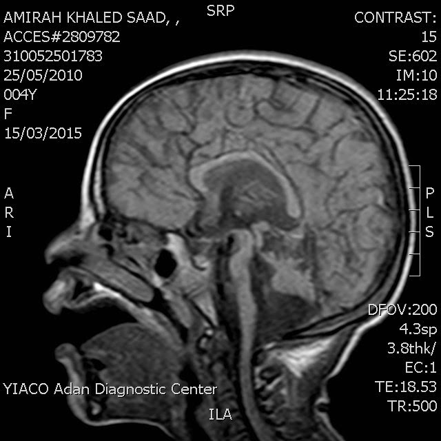
T1W – Sagittal & axial scans
T2W – Axial & coronal scans
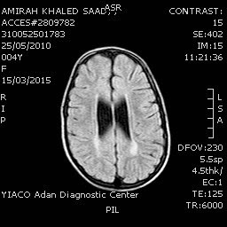
FLAIR – Axial scans
FINDINGS:
-Revealed:
• Corpus callosal atrophy.
• Brain stem atrophy.
• Severe degree of atrophy of the cerebellum.
• Dilated lateral and third ventricles.
• Mild peri ventricular white matter ischemic changes mainly around occipital horns of lateral ventricles.
• Fourth ventricle maintains its normal size but with widening of the area of outlet foramina.
-The rest of the brain Parenchyma shows normal grey white matter differentiation and signal intensities. No focal mass lesion is seen.
-Basal cisterns, cortical sulci and gyri are normal in appearance.
DISCUSSION& IMPRESSION:
• On the light of findings seen in this case; the whole land mark is severe atrophy of the cerebellum.
• Atrophy of the cerebellum is associated with many genetic conditions.
• Non genetic causes include chronic severe alcohol intake, para neoplastic syndromes, drugs like Phenytoin.
• The nomenclature of genetic disorders associated with cerebellar atrophy is complex. Most are classified by the chromosomal location and pattern of inheritance. In many, a specific gene mutation or defective protein has been found. In the past, these disorders were referred to as cerebellar degeneration, spinocerebellar degeneration and olivopontocerebellar atrophy (OPCA) depending on whether the cerebellum, spinal cord, brainstem or a combination of any of the above affected.
Reported By : DR.MOHAMMED MOHYELDIN
Consultant-Radiologist
Report Status : Validated / Validated By : Dr.Mohammed Mohyeldin
No comments:
Post a Comment